From sun spots to post-inflammatory discoloration, consumers continue to seek products that promote brighter, more even skin tone. In one study, participants with hyperpigmentation reported a willingness to spend over $80 per month for a 50% improvement in their skin condition (Jafry et al., 2024). However, claims like “brightening” or “tone-evening” carry limited weight without scientific backing. Products and ingredients that deliver measurable results are those best positioned for success among science-minded consumers. To address this need, Genermarkers offers an in vitro melanin inhibition assay for evaluating pigmentation science and UV-induced melanogenesis.
The Biology Behind Hyperpigmentation
Each step in the hyperpigmentation process—from the activation of melanogenic enzymes to the signaling pathways that regulate melanosome production and transfer—can be measured and quantified. Understanding and targeting these biological processes is the key to successful anti-hyperpigmentation formulation.
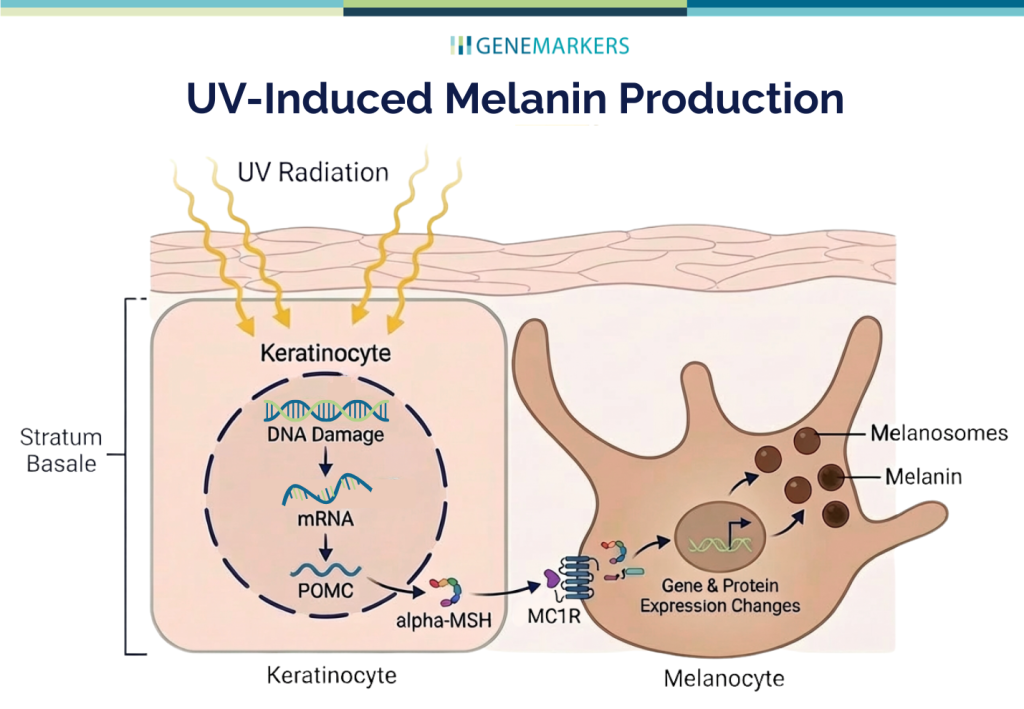
Melanin Production (Melanogenesis)
When UV radiation reaches the keratinocytes in the stratum basale of the epidermis, it induces DNA damage, triggering the production and release of alpha-melanocyte-stimulating hormone (ɑ-MSH). This signaling molecule binds to the melanocortin 1 receptor (MC1R) on neighboring melanocytes, initiating a cascade of intracellular responses. As a result, key melanogenic enzymes, including tyrosinase, are upregulated, leading to increased melanin synthesis within specialized organelles known as melanosomes. While complex, each step in this pathway is a measurable molecular event. Measuring and quantifying the molecular processes involved in melanogenesis can help inform product development and validate the efficacy of anti-hyperpigmentation formulations and ingredients.
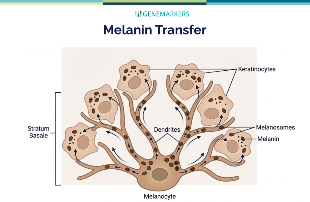
Melanin Transfer
Melanin transfer also plays a significant role in skin pigmentation. During melanin transfer, pigment-filled melanosomes are transferred from melanocytes to keratinocytes. Once inside the keratinocyte, the melanosomes localize around the nucleus to help protect DNA from UV damage. While this serves an important protective function, repeated UV exposure can disrupt this balance, leading to excessive or uneven pigment distribution.
Biomarker analysis measures and quantifies changes in key genes and proteins involved in melanogenesis and melanin transfer. This insight enables product formulators and ingredient developers to create effective hyperpigmentation solutions based on biological data—the key to success in an increasingly competitive and science-driven market.
From Brightening Claims to Biological Targets
Today’s most effective hyperpigmentation ingredients and formulations are designed to influence the molecular pathways involved in pigmentation. As our understanding of melanogenesis and hyperpigmentation advances, formulation strategies are shifting from broad aesthetic claims toward targeted biological intervention.
While traditional evaluation methods, such as visual grading or consumer perception studies, offer valuable insights, they provide limited visibility into biological performance. As hyperpigmentation research becomes increasingly mechanism-driven, formulators need tools that go beyond surface-level observations to provide measurable, reproducible data. Melanin inhibition testing, using primary human melanocytes, fills this gap by providing a direct, quantitative assessment of melanin production under controlled conditions.
Advancing Hyperpigmentation Research with Genemarkers
To meet the growing demand for biologically relevant testing, Genemarkers has developed an in vitro melanin inhibition assay for evaluating UV-induced melanogenesis and pigmentation.
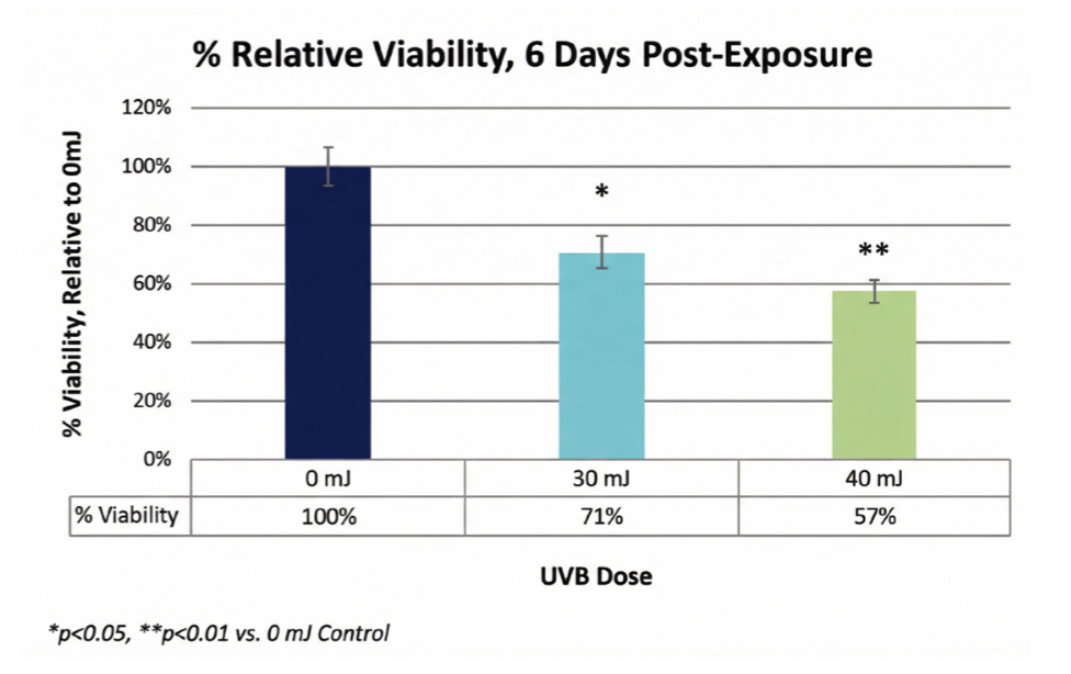
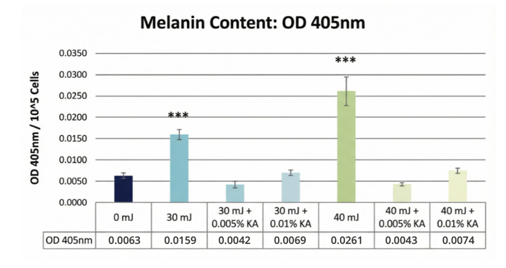
Using normal human primary melanocytes, the model induces melanin production through controlled UVB exposure. Validation studies demonstrate that UVB doses of 30 and 40 mJ/cm² significantly increased melanin content compared to non-irradiated controls. As expected, UV exposure produced a dose-dependent reduction in cell viability, reflecting real biological stress conditions.
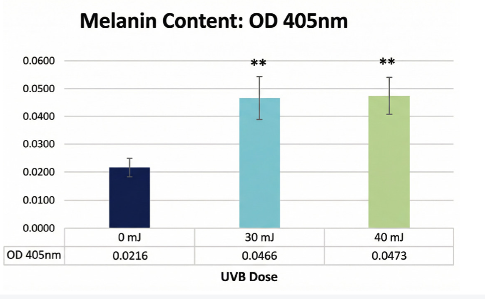
To confirm assay sensitivity and further validate the model, kojic acid was used as a reference inhibitor. When applied prior to UV exposure and maintained throughout the study period, treated cells exhibited melanin levels comparable to non-irradiated controls, demonstrating effective suppression of UV-induced pigmentation. Using this model, study sponsors can evaluate how ingredients or formulations affect pigmentation and UV-induced melanogenesis.
Applications of the testing assay include:
- Screening novel compounds for anti-pigmentation activity
- Benchmarking against known brightening agents
- Supporting claims related to skin tone improvement
- Evaluating dose-response relationships
- Understanding mechanisms of action
To take this data to the next level, Genemarkers has developed a proprietary, off-the-shelf, qPCR panel that contains 50 biomarker genes that play a role in melanogenesis and pigmentation.
Sources:
- Jafry M, Guan LL, Mohammad TF. A practical guide to over-the-counter treatments for hyperpigmentation. JEADV Clin Pract. 2024;3:433–447. https://doi.org/10.1002/jvc2.385